How Does Facial Fat Change With Age? Why the Face Can Look Hollow and Saggy at the Same Time | Reva Clinic
When we talk about facial aging, most people think first about collagen loss, wrinkles, or skin laxity.
However, facial aging does not happen only at the skin level.
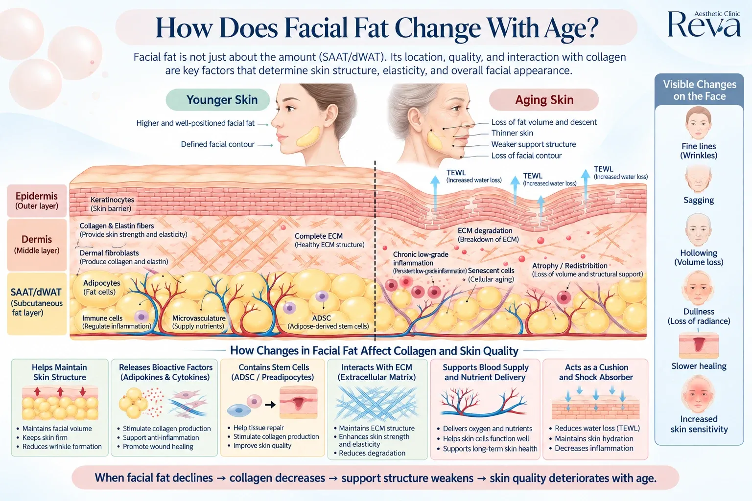
One of the most important structures that changes with age is facial fat. It contributes to facial volume, contour, skin smoothness, and structural support.
More importantly, facial fat does not simply disappear evenly as we age.
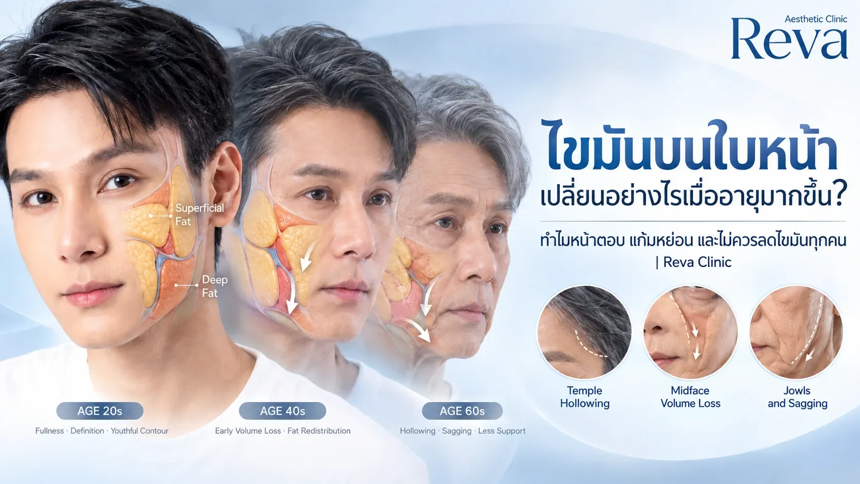
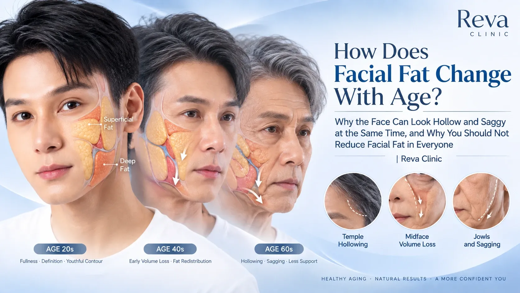
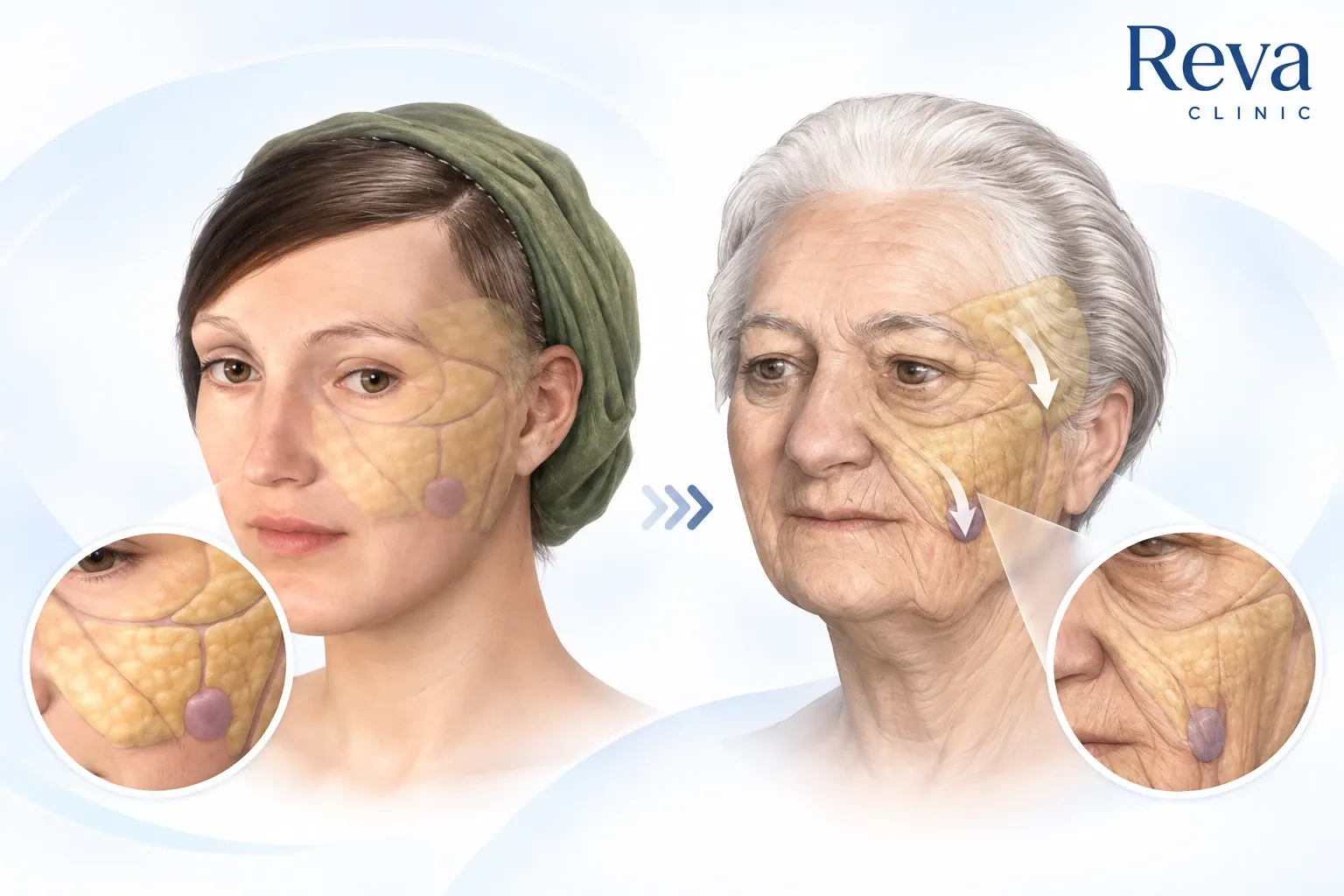
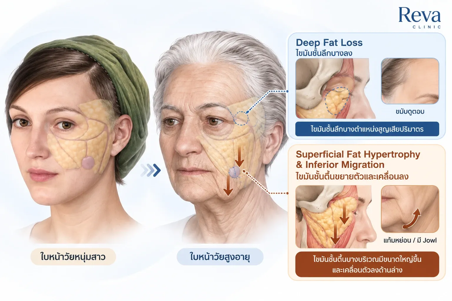
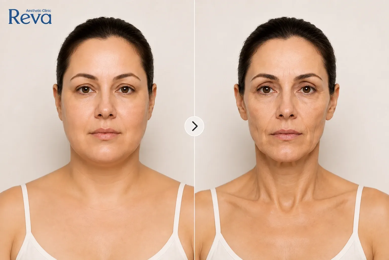
Some areas may develop fat atrophy, leading to hollow temples or flatter cheeks. At the same time, fat in other areas may enlarge or undergo fat redistribution, contributing to deeper folds, lower-face heaviness, and jowls.
This explains why the same person can look both hollow and saggy at the same time.
Therefore, when treating the aging face, the first question should not simply be:
“How can we reduce facial fat?”
A better question is:
“How is the fat in each area and layer of the face changing?”
Key Takeaways
Facial fat does more than create fullness. It also contributes to facial structure, support, and communication with surrounding tissues.
Adipose tissue includes different biological types, such as White Adipose Tissue (WAT) and Brown Adipose Tissue (BAT).
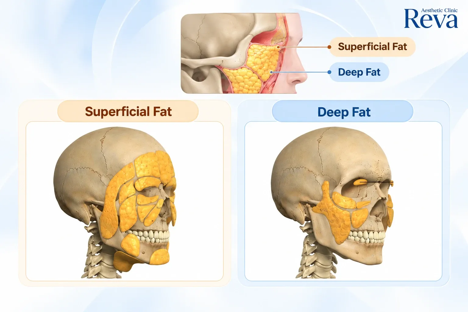
Anatomically, facial fat can also be divided into Superficial Fat and Deep Fat.
With age, some deep fat compartments may lose volume, while some superficial fat compartments may enlarge or shift in position.
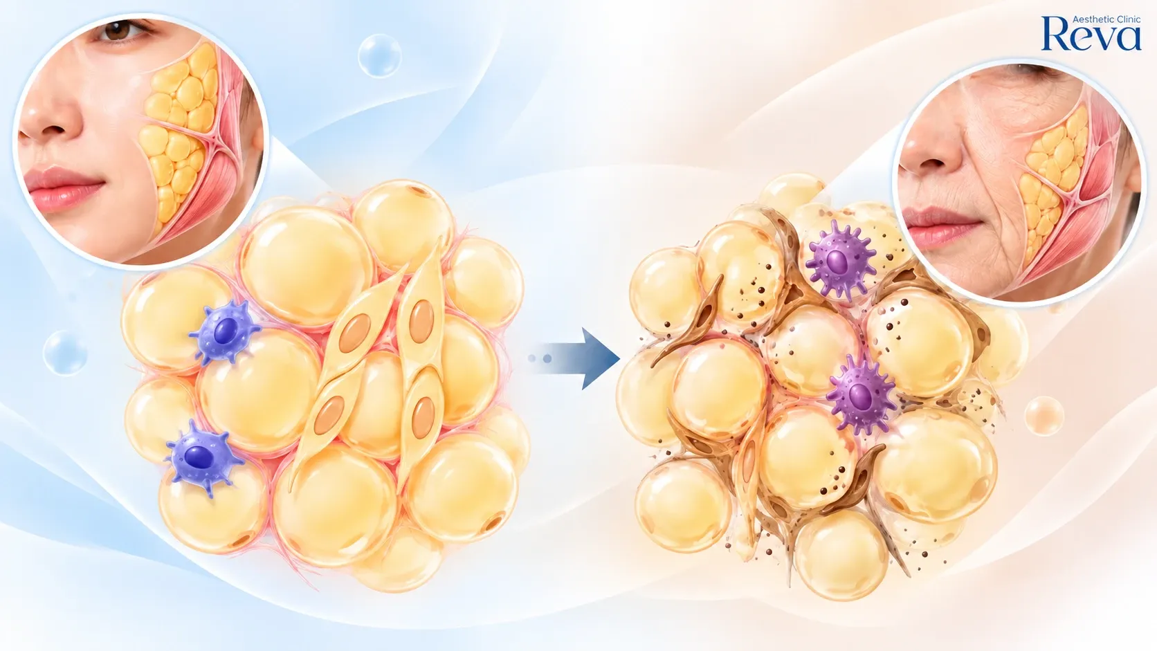
Fat cells themselves also age, with reduced regenerative capacity, increased inflammation, and changes in brown and beige adipose tissue.
Fibrous septa, the connective tissue partitions that organize and support fat compartments, may also change with age.
Facial rejuvenation should therefore consider whether each area should be Reduced, Preserved, Restored, or Tightened, rather than trying to reduce fat everywhere.
Facial Fat Does More Than Make the Face Look Full
Facial fat is often viewed simply as something that makes the face look larger.
This can lead to the assumption that:
“The more facial fat we remove, the slimmer and younger the face will look.”
In reality, adipose tissue has many functions beyond energy storage.
It provides mechanical cushioning, fills spaces between anatomical structures, contributes to facial volume and contour, and helps create gliding planes that allow different tissue layers to move over one another.
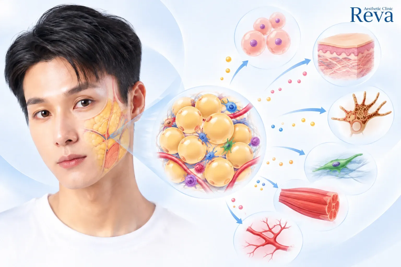
Adipose tissue is also biologically active. It releases adipokines, which are signaling molecules produced by fat tissue, as well as other factors involved in metabolism, inflammation, immune function, and communication with surrounding cells.
Facial fat should therefore not simply be considered unwanted tissue beneath the skin.
It is a living and biologically active component of facial structure.
What Is the Difference Between White Fat and Brown Fat?
When we use the word “fat,” we are not actually referring to only one type of adipose tissue.
There are several types of fat cells, but two of the best known are:
White Adipose Tissue (WAT)
and
Brown Adipose Tissue (BAT)
They have different biological functions.
What Is White Adipose Tissue (WAT)?
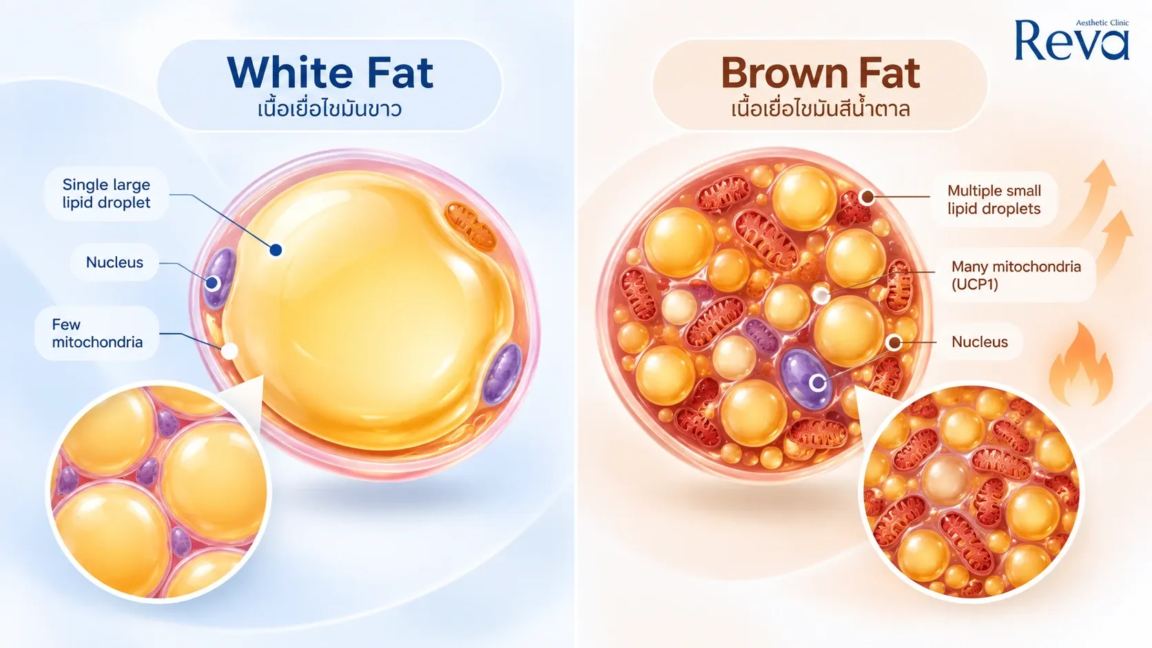
White Adipose Tissue, or WAT, is the predominant type of adipose tissue in the human body.
One of its main functions is to store excess energy in the form of triglycerides and release that energy when needed.
However, white fat is not simply an energy-storage compartment.
It also functions as an active endocrine and signaling tissue, releasing adipokines and other molecules that influence metabolism, inflammation, immune function, and surrounding cells.
In the face and skin, white adipose tissue also contributes to:
Therefore, white fat should not automatically be considered “unwanted fat.”
What matters is how much is present, where it is located, and how healthy the tissue is.
What Is Brown Adipose Tissue (BAT)?
Brown Adipose Tissue, or BAT, behaves differently from white fat.
Instead of primarily storing energy, brown fat is specialized for thermogenesis, which means using energy to generate heat.
Brown adipocytes contain large numbers of mitochondria, the structures inside cells involved in energy production. This allows brown fat to consume energy and generate heat.
In simple terms:
White Fat → mainly stores energy
Brown Fat → mainly uses energy to produce heat
This does not mean that white fat is “bad” and brown fat is “good.”
They simply have different biological functions.
What Is Beige Fat?
There is another interesting group known as Beige Adipocytes.
Under certain biological conditions, beige fat cells can develop thermogenic characteristics similar to brown fat.
This ability of adipose tissue to change its characteristics and biological behavior is part of what is known as:
Adipocyte Plasticity — the ability of fat cells to adapt and change their functional characteristics.
What Happens to Brown and Beige Fat as We Age?
Aging does not simply determine whether we have “more fat” or “less fat.”
The biology of adipose tissue itself also changes.
With aging, studies have reported changes involving Brown Adipose Tissue and Beige Adipocytes, together with broader changes in adipose metabolism.
At the same time, Adipocyte Progenitor Cells, the precursor cells that can develop into mature fat cells, may become dysfunctional with age.
This can contribute to:
Reduced adipogenesis, or formation of new fat cells
Reduced adipose tissue renewal
Metabolic imbalance
Abnormal fat distribution
Adipose tissue dysfunction
References: Front Endocrinol (Lausanne). 2026;17:1821542; Cell Death Dis. 2022;13(4):300.
Therefore, fat aging does not simply mean losing fat.
The amount, location, metabolism, regenerative capacity, and biological environment of adipose tissue can all change with age.
White Fat vs Brown Fat Is Not the Same as Superficial Fat vs Deep Fat
This distinction is important.
White Fat vs Brown Fat describes the biological type and function of adipose tissue.
In contrast,
Superficial Fat vs Deep Fat describes the anatomical location of facial fat.
These are two different ways of classifying adipose tissue.
Therefore:
Superficial Fat ≠ White Fat
and
Deep Fat ≠ Brown Fat
Understanding this distinction makes it easier to understand how different aspects of facial fat can change with age.
How Many Layers of Facial Fat Are There?
For easier understanding, facial fat can broadly be considered in two major anatomical groups.
1. Superficial Fat
Superficial Fat is located relatively close to the skin.
Its fat lobules are generally smaller and more tightly packed, with fibrous structures running between them and helping anchor the tissue.
Because of this, superficial fat plays an important role in surface contour, or how smooth and well-defined the face appears from the outside.
When this layer changes, we may notice downward displacement of the cheeks, uneven contours, or reduced jawline definition.
2. Deep Fat
Deep Fat lies deeper within the face and generally consists of larger, flatter fat lobules.
It contributes significantly to facial volume, and its thickness can vary according to the anatomical area, age, sex, and changes in body weight.
The distinction between these two layers is important because:
Superficial and deep facial fat do not necessarily age in the same way.
Does Facial Fat Really Disappear as We Age?
Partly, but that is not the whole story.
Facial aging involves more than simply fat loss.
It also involves:
Fat Redistribution — Changes in the Position and Distribution of Facial Fat
With aging, some deep fat compartments may deflate or undergo atrophy.
At the same time, some superficial fat compartments may become relatively enlarged or undergo changes in distribution.
References: Plast Reconstr Surg Glob Open. 2014;1(9):e92; Skin Res Technol. 2022;28(6):872–876.
As a result, aging does not necessarily make the entire face uniformly thinner.
The same face may develop:
Hollow temples
Loss of midface fullness
More prominent nasolabial folds
Downward displacement of the cheeks
Reduced jawline definition
Jowls
all at the same time.
Why Can the Face Look Hollow and Saggy at the Same Time?
This is one of the patterns that can occur with facial aging.
Some deep fat compartments, particularly around the temples and midface, may lose volume.
At the same time, certain superficial fat compartments may undergo hypertrophy, meaning enlargement of fat cells, together with inferior migration, or downward displacement.
The visible result can therefore be:
“Less volume in the upper face, but more heaviness in the lower face.”
This is why seeing a jowl and immediately concluding:
“There is too much fat, so we should reduce it.”
may not tell the whole story.
Fat Itself Can Age
In addition to changes in fat volume and location, the adipose tissue itself can undergo age-related changes.
Healthy adipose tissue has regenerative capacity and can maintain tissue homeostasis.
With age, several changes may occur, including:
Reduced regenerative capacity
Enlargement of fat cells
Increased inflammation
Changes in brown and beige adipose tissue
Dysfunction of adipocyte progenitor cells
Reduced formation of new fat cells
Abnormal fat distribution
References: Aesthetic Plast Surg. 2021;45:151–163; Cell Death Dis. 2022;13(4):300; Front Endocrinol (Lausanne). 2026;17:1821542.
Therefore:
Having “more fat” is not the same as having healthy adipose tissue.
What Does Facial Fat Have to Do With Collagen and Skin Quality?
The skin and the fat beneath it do not function completely independently.
Adipocytes, or fat cells, can release signaling molecules that influence surrounding cells.
Research suggests that factors derived from adipose tissue may influence fibroblasts, the cells involved in producing collagen and other components of the skin’s structural matrix.
Research has also investigated Extracellular Vesicles (EVs), tiny particles used for cell-to-cell communication.
In experimental models, adipose-derived EVs have been associated with changes in skin thickness, collagen production, and cellular senescence, or the state in which aging cells stop dividing normally but remain biologically active.
This supports an increasingly important concept:
Skin aging and fat aging may be interconnected rather than completely separate processes.
What Is Dermal White Adipose Tissue (dWAT)?
In addition to superficial and deep facial fat, another interesting component is:
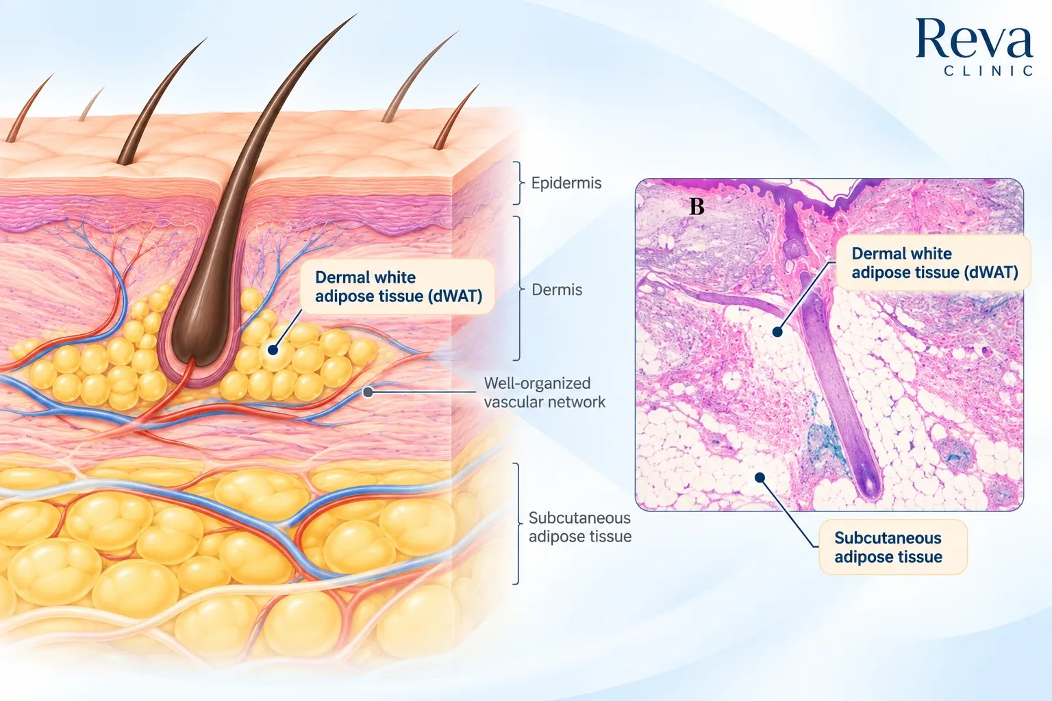
Dermal White Adipose Tissue, or dWAT
This refers to white adipose tissue located within or closely associated with the dermis.
dWAT has been associated with several biological processes, including:
Hair follicle cycling
Wound healing
Thermoregulation
Immune defense
Cutaneous fibrosis
References: Aging (Albany NY). 2016;8(7):1457–1469; J Cosmet Dermatol. 2026;25(1):e70671.
Fatty acids from dWAT can also provide metabolic substrates for fibroblasts, while dWAT appears to contribute to Extracellular Matrix (ECM) homeostasis, meaning the maintenance of the structural environment surrounding cells, as well as the mechanical properties of the dermis.
Reference: Front Physiol. 2024;15:1346612.
This is another reason why facial aging should not always be considered a skin-only problem.
How Can Aging Fat Affect Skin Quality?
Changes in adipose tissue may also affect the surrounding skin.
Research into dermal fat infiltration, where adipose tissue extends into or interacts differently with the dermal layer, has linked these changes with structural alterations and reduced dermal elasticity.
Mechanistic studies have also investigated MMP-9 and palmitic acid.
Under experimental conditions, these pathways may be associated with elastic fiber degradation, reduced collagen and elastin expression, and increased MMP-1 expression.
Reference: Skin Res Technol. 2022;28(6):872–876.
Therefore, age-related changes in facial fat may affect more than whether the face looks “fatter” or “thinner.”
They may also be associated with changes in the quality of the surrounding tissues.
What Are Fibrous Septa, and Why Does Facial Fat Stay in Compartments?
Facial fat is not one large layer that moves freely underneath the skin.
It is divided into smaller compartments by structures known as:
Fibrous Septa — connective tissue partitions that separate fat into smaller lobules.
These structures help organize facial fat and connect it with surrounding tissues.
Another important structure is the:
Retaining Ligament — a strong fibrous attachment connecting deeper structures, such as bone or deep facial fascia, with more superficial layers.
There is also the:
Retinacula Cutis — smaller fibrous structures that connect and support the skin and underlying tissues.
A simple way to think about it is:
Facial fat does not float freely underneath the skin. It is organized and supported by a network of fibrous tissue.
The Fibrous Support Around Facial Fat Also Changes With Age
Aging does not affect only fat cells or fat volume.
The Fibrous Septa and Retinacula Cutis that organize and support facial fat can also undergo age-related changes.
A reduction in connective tissue within fat pads may contribute to a greater tendency for the tissue to sag.
Reference: Kwon et al. 2026. doi:10.1007/s12565-026-00936-8.
This may help explain why someone can feel:
“My face is starting to drop.”
even without a major increase in facial fat.
The problem may involve changes in structural support as well as changes in fat volume.
Why Reducing More Facial Fat Is Not Always Better
Consider someone with:
Hollow temples + midface volume loss + deeper nasolabial folds + jowls
If we look only at the jowls, it may seem logical to conclude:
“There is too much fat. We should reduce it.”
But when the entire face is assessed, some deep fat compartments may already be losing volume while superficial fat in other areas is changing position.
Aggressive fat reduction in someone who already has facial volume loss may therefore not always be the appropriate goal.
A more useful approach is to determine:
Which areas should be reduced, which should be preserved, and which have lost volume?
How Can We Approach Aging Facial Fat?
An important concept in facial rejuvenation is that treatment should not focus solely on removing fat.
At least three factors should be considered:
Fat Quantity — How much fat is present?
Fat Quality — What is the biological condition of the adipose tissue?
Fibrous Support — How well are the surrounding supporting structures functioning?
Treatment can then be selected according to the problem and tissue layer of each individual.
Can Hyaluronic Acid (HA) Do More Than Restore Volume?
Hyaluronic Acid, or HA, is commonly used in injectable fillers to replace lost facial volume.
Experimental research has also suggested that HA may influence the adipose microenvironment.
In experimental models, HA has been associated with maintained adipogenic capacity, improved cell adherence during adipocyte differentiation, increased lipid accumulation and adiponectin secretion, and reduced lipolysis.
Reference: J Cosmet Dermatol. 2021;20(5):1474–1482.
However, this was experimental research.
It should not be interpreted to mean that injecting HA into a patient directly “regenerates facial fat” or creates new fat cells.
Clinically, the established role of HA remains primarily related to restoring volume and contour when appropriately indicated.
Collagen Biostimulators May Affect More Than Collagen Alone
Collagen Biostimulators are another area of interest because the tissue response may extend beyond the superficial dermis.
For example, studies involving Polycaprolactone (PCL) have investigated interactions with M2 macrophages, immune cells associated with tissue repair, as well as fibroblast activation.
Studies have reported increases in Type I collagen, Type III collagen, and elastin, together with changes toward thicker and stronger fibrous septa.
References: Stem Cell Res Ther. 2023;14(1):321; Aesthet Surg J. 2025;45(5):514–524.
This expands the concept of biostimulation beyond simply:
“Making more collagen in the skin.”
Researchers are increasingly investigating how biostimulatory materials interact with the broader tissue environment.
PLLA, or Poly-L-lactic Acid, is best known as a collagen biostimulator.
However, mechanistic research has also investigated interactions between PLLA and Adipose-Derived Stem Cells (ADSCs), which are stem/progenitor cells derived from adipose tissue.
Pathways involving NRF2, C/EBPα, and PPARγ have been investigated in relation to adipogenesis and lipogenesis.
Reference: Polymers (Basel). 2025;17(13):1826.
However, this does not mean that PLLA injections should be considered a treatment for creating new facial fat.
These findings are mechanistic and should not be directly translated into a clinical claim that PLLA regenerates facial fat in humans.
For patients with changes in skin quality and tissue structure, collagen biostimulators may be considered depending on individual indications and physician assessment.
Does Monopolar RF Cause Facial Fat Loss?
One common concern before a lifting treatment is:
“Will an energy-based lifting treatment make my face look hollow?”
Monopolar RF (Monopolar Radiofrequency) uses radiofrequency energy to generate controlled heat within tissue.
However, the treatment goal does not necessarily have to be:
“Destroy as much fat as possible.”
Research into Mild Hyperthermia, or lower-temperature controlled heating below 43°C, has investigated biological effects involving angiogenesis, adipose-derived stem cell activity, and adipose tissue metabolism.
References: Life Med. 2022;1:78–80; Biomater Res. 2023;27:122.
Therefore, particularly in someone who already has facial volume loss, Monopolar RF treatment should consider both tissue laxity and the volume that should be preserved.
At Reva Clinic, Program Oligio X uses Monopolar RF technology, with treatment planning individualized according to facial structure and the concerns in different areas.
For patients experiencing facial volume loss or laxity after significant weight loss, read more about:
Program Oligio X for Post-GLP-1 Face and the X Layer Protocol
Why Can the Face Look Older After Significant Weight Loss?
People who lose a significant amount of weight may notice that their face appears more hollow or lax.
The term “Ozempic Face” is commonly used online. More broadly, these changes can be considered Post-GLP-1 Facial Changes, particularly when significant weight loss occurs during treatment with GLP-1 medications.
Changes may include loss of subcutaneous volume around the cheeks, temples, periorbital area, and neck.
Loss of lean mass, including muscle, may also contribute to reduced facial support and make laxity more visible.
However, whether semaglutide has a specific direct lipolytic effect on facial fat remains unclear.
Reference: doi:10.1002/der2.70003.
Therefore, Post-GLP-1 facial changes should not simply be described as “the medication melting facial fat.”
Should Your Face Be Reduced, Restored, Preserved, or Lifted?
Instead of starting with questions such as:
“Which device is the strongest?”
“How many shots do I need?”
or
“How much fat can we remove?”
a more useful question is:
Which tissue layer is actually causing the problem?
If there is Deep Fat Atrophy
→ the goal may be Restore — replacing lost volume.
If facial fat is still present in an appropriate amount and position
→ the goal may be Preserve — maintaining valuable facial volume.
If there is genuine Localized Fat
→ the goal may be Reduce — selectively reducing the appropriate area.
If the main concern is Skin Laxity
→ the goal may be Tighten / Lift — improving tissue firmness and support.
If tissue quality has changed
→ Biostimulation may be considered when appropriately indicated.
The same person may therefore require different approaches in different parts of the face.
Why Personalized Facial Design Matters
Every face has a different amount and distribution of fat, different tissue thickness, different supporting structures, and a different pattern of aging.
Facial rejuvenation should therefore not begin only with:
“How many shots?”
or
“How much energy?”
It should begin with:
“Which tissue layer is changing, and what do we want to achieve in that layer?”
This is the concept behind Personalized Facial Design.
The goal is not to make every layer thinner or tighter.
The goal is to maintain balance between facial volume, contour, tissue support, and skin quality.
Summary: Facial Rejuvenation Is About Balance, Not Simply Removing Fat
Facial aging does not occur in only one layer.
With age, several changes may happen together:
Fat Redistribution — changes in where facial fat is located
Deep Fat Atrophy — loss of deeper facial volume
Fat Cell Aging — age-related changes in fat cell function
Changes in Brown and Beige Fat — age-related changes in thermogenic adipose tissue
Fibrous Support Aging — changes in the structures supporting facial tissues
along with changes in skin and collagen.
Therefore, the goal of facial rejuvenation should not simply be:
“Remove as much facial fat as possible.”
Instead, the aim should be:
Appropriate fat quantity + healthy tissue quality + appropriate structural support
In simpler terms:
Facial fat should be present in the right amount, in the right place, with adequate surrounding support.
Understanding which facial layer is changing allows treatment to be planned more precisely:
Reduce what is excessive
Preserve what is valuable
Restore what has been lost
Tighten what has become lax
rather than applying the same treatment pattern to every area of the face.
References
Plast Reconstr Surg Glob Open. 2014;1(9):e92.
Skin Res Technol. 2022;28(6):872–876.
Aesthetic Plast Surg. 2021;45:151–163.
Cell Death Dis. 2022;13(4):300.
Front Endocrinol (Lausanne). 2026;17:1821542.
Aging (Albany NY). 2016;8(7):1457–1469.
J Cosmet Dermatol. 2026;25(1):e70671.
Front Physiol. 2024;15:1346612.
Kwon et al. 2026. doi:10.1007/s12565-026-00936-8.
J Cosmet Dermatol. 2021;20(5):1474–1482.
Stem Cell Res Ther. 2023;14(1):321.
Aesthet Surg J. 2025;45(5):514–524.
Polymers (Basel). 2025;17(13):1826.
Life Med. 2022;1:78–80.
Biomater Res. 2023;27:122.
doi:10.1002/der2.70003.