Is Subcision Still Necessary for Acne Scar Treatment in 2026? Latest Evidence, New Techniques, and Evolving Treatment Strategies
Dr. Songsak Suksantilap (Dr. Bank)
August 6, 2026 · Reading time 15 minutes
Share
Is Subcision Still Necessary for Acne Scar Treatment in 2026?
Latest Evidence, New Techniques, and Evolving Treatment Strategies
With the emergence of advanced lasers, RF microneedling, and collagen biostimulators, is subcision still necessary for acne scar treatment? This article explores the latest evidence, updated techniques, potential complications, and combination treatment strategies to help you choose the most effective approach for each type of acne scar.
1. What Is Subcision?
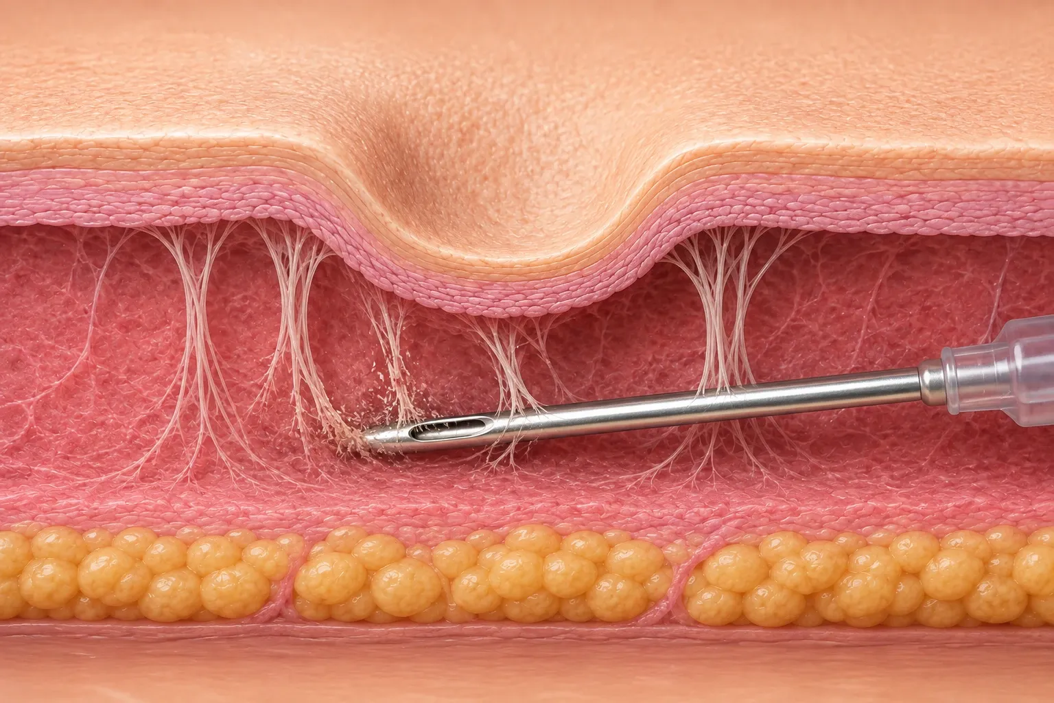
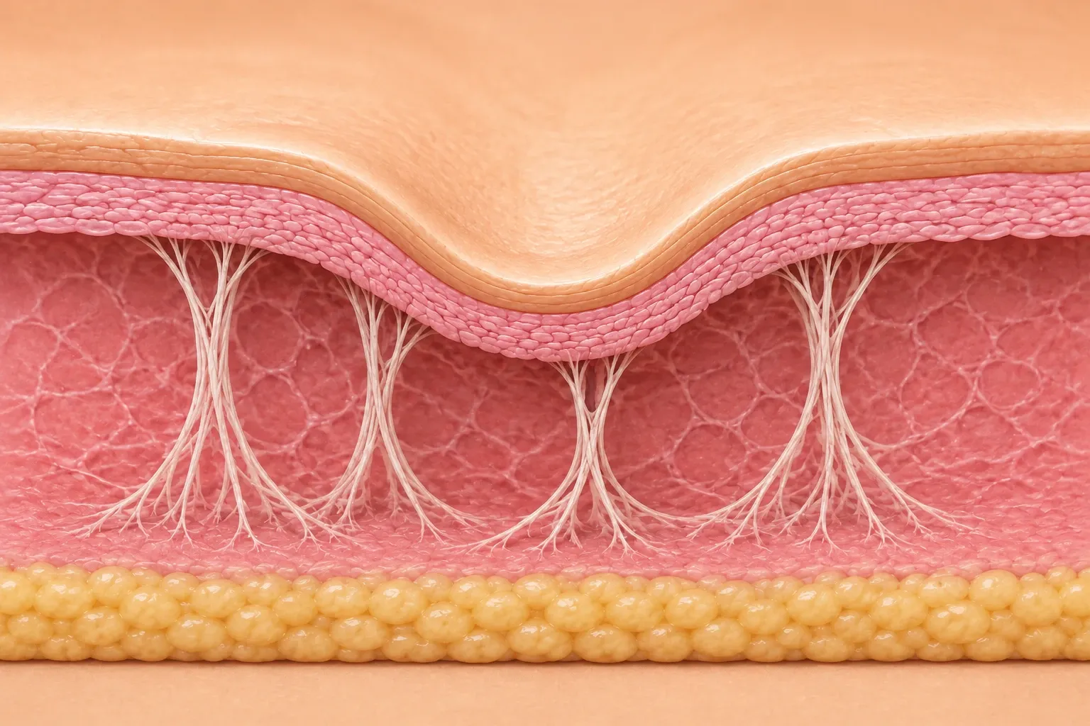
Subcision is a minimally invasive procedure designed to treat tethered atrophic acne scars by releasing the fibrous bands (fibrous tethering) beneath the skin.
During the procedure, a physician inserts a sharp needle or blunt cannula beneath the scar to carefully release these fibrotic attachments. Once the tethering is released, the depressed skin can elevate, while the controlled wound-healing response stimulates new collagen formation and tissue remodeling, leading to gradual improvement in scar appearance.
Objectives of Subcision
Release fibrotic bands beneath the scar
Eliminate pathological skin tethering
Stimulate collagen remodeling
Enhance the effectiveness of combination treatments such as lasers, RF microneedling, fillers, and biostimulators
Clinical Insight
Subcision does not directly treat the scar itself. Instead, it addresses the underlying fibrous tethering, which is often the primary cause of depressed acne scar
2. Which Acne Scars Benefit Most from Subcision?
One of the most common misconceptions is that all acne scars can be treated with subcision.
In reality, subcision is effective only when fibrous tethering is present. Because the procedure works by releasing fibrotic attachments beneath the skin, its success depends more on scar morphology than on scar depth alone.
Rolling Scar
⭐⭐Best Indication
Rolling scars are considered the ideal indication for subcision.
These scars typically appear as broad, shallow depressions with poorly defined borders, creating a wave-like contour across the skin. The underlying cause is usually fibrous septae that tether the dermis to deeper tissues.
Once these fibrous bands are released, several beneficial effects occur:
Immediate elevation of the depressed skin
Reduction of tethering forces
Stimulation of collagen remodeling
Improved response to subsequent laser or RF microneedling treatments
Clinical Pearl
If the underlying tethering is not released, repeated laser treatments alone may provide only limited improvement because the primary mechanical cause of the scar remains unchanged.
Rolling Scar
Boxcar Scar
⭐Only When Tethering Is Present
Not every boxcar scar requires subcision.
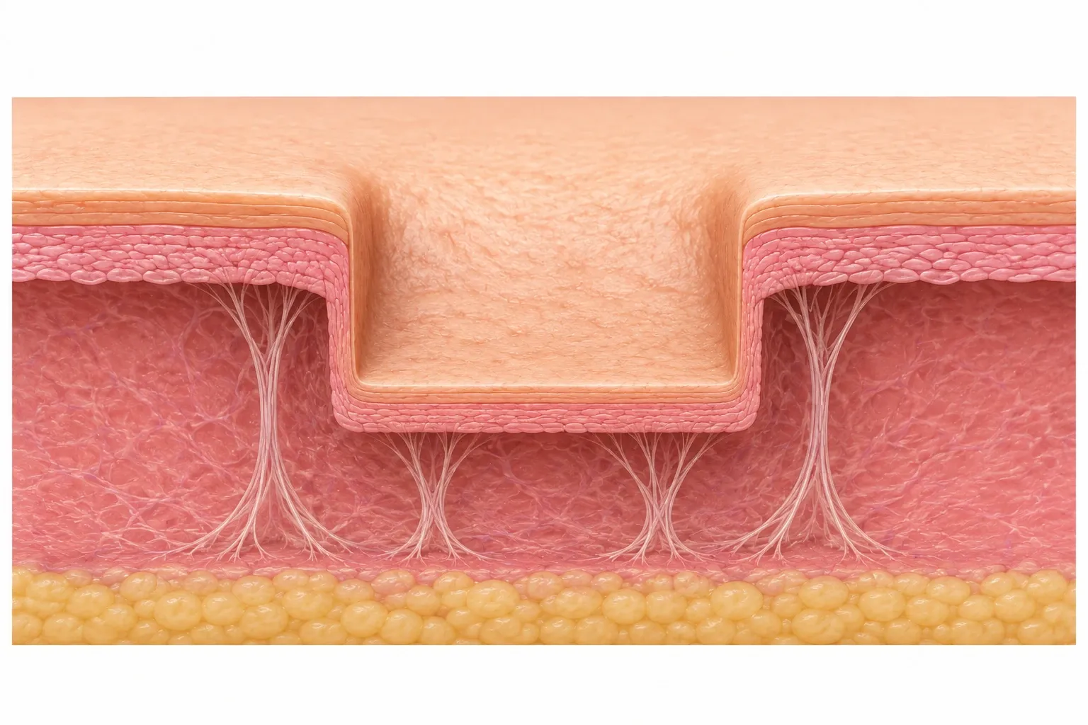
Boxcar scars have sharply defined borders and a relatively flat base. Some are associated with fibrous tethering, while others are caused primarily by tissue loss without significant tethering.
When tethering is present, subcision may help by:
Releasing fibrous attachments
Reducing scar depth
Improving outcomes when combined with laser resurfacing
However, if no tethering exists, subcision usually offers limited benefit. Alternative treatments may include:
Fractional CO₂ Laser
Er:YAG Laser
RF Microneedling
TCA CROSS (selected cases)
Boxcar Scar
Ice Pick Scar
❌ Not an Indication for Subcision
Ice pick scars are not suitable for subcision.
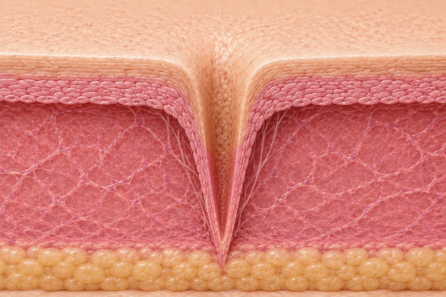
These scars are narrow, deep, and vertically oriented, resulting primarily from loss of tissue rather than fibrous tethering.
Because there is little or no fibrous band to release, subcision generally produces minimal improvement.
More appropriate treatments include:
TCA CROSS
Punch Excision
Punch Elevation
Ablative Laser Resurfacing
Ice Pick Scar
Subcision is designed to release the underlying fibrous tethering rather than the acne scar itself. As a result, successful treatment depends on careful assessment of scar morphology and the presence of fibrotic attachments, rather than scar depth alone.
3. How Has the Treatment Philosophy Changed in 2026?
Historically, subcision was often performed using an aggressive approach, with the goal of releasing as many fibrous attachments as possible to eliminate skin tethering.
Today, however, the treatment philosophy has shifted toward Precision Subcision—a targeted approach that focuses on releasing only the pathological fibrous bands responsible for acne scar tethering while preserving surrounding healthy tissue whenever possible.
This evolution reflects a better understanding of facial anatomy and wound healing. Rather than performing extensive undermining, modern subcision aims to maximize treatment effectiveness while minimizing unnecessary tissue trauma.
Excessive or overly aggressive subcision may increase tissue injury, leading to a higher risk of hematoma, prolonged swelling and bruising, new fibrosis, and longer recovery times. In some cases, excessive tissue disruption may also contribute to contour irregularities or treatment-related scarring.
As a result, successful subcision is no longer defined by how much tissue is released, but by how precisely the pathological fibrous tethering is identified and treated. Proper patient selection, accurate treatment depth, appropriate instrument choice, and meticulous surgical technique all play critical roles in achieving optimal clinical outcomes.
Goals of Precision Subcision
Release only the fibrous bands responsible for skin tethering
Minimize unnecessary tissue trauma
Reduce the risk of hematoma formation
Decrease swelling, bruising, and downtime
Preserve normal tissue and facial support structures
Clinical Pearl
A successful subcision is not about releasing more tissue—it is about releasing the right tissue. Modern subcision emphasizes targeted treatment of pathological fibrous tethering while preserving healthy tissue, forming the foundation of today's Precision Subcision concept.
4. High-Risk Anatomical Areas for Subcision
Although subcision is generally considered a safe procedure when performed by an experienced physician, certain areas of the face carry a higher risk of complications, including hypertrophic scarring, vascular injury, and prolonged swelling. Careful anatomical assessment is therefore essential before treatment.
Areas Requiring Extra Caution
Perioral Area (Around the Mouth)
The skin around the mouth is highly mobile due to constant facial movement during speaking, eating, and smiling. This repetitive mechanical tension may increase the risk of hypertrophic scar formation and prolonged healing.
Periorbital Area (Around the Eyes)
The periorbital region has extremely thin skin and contains numerous blood vessels, nerves, and delicate anatomical structures. Subcision in this area requires meticulous technique and a thorough understanding of facial anatomy.
Perinasal Area (Around the Nose)
The perinasal region contains dense fibrous attachments and a rich vascular network. Procedures performed in this area require particular caution to minimize tissue trauma and avoid injury to important anatomical structures.
Why Are These Areas Considered High Risk?
Performing subcision in these regions with inappropriate technique or excessive tissue release may increase the risk of:
Hypertrophic scar formation
Hematoma
Excessive swelling and bruising
Injury to blood vessels or other critical facial structures
For this reason, successful subcision depends not only on choosing the appropriate instrument but also on accurately evaluating scar type, fibrous tethering, anatomical location, and treatment depth. The procedure should be performed by a physician with a comprehensive understanding of facial anatomy and extensive experience in acne scar management.
Clinical Pearl
Safe subcision is guided by anatomy, not force. Careful patient assessment and precise technique are essential for maximizing treatment outcomes while minimizing the risk of complications.
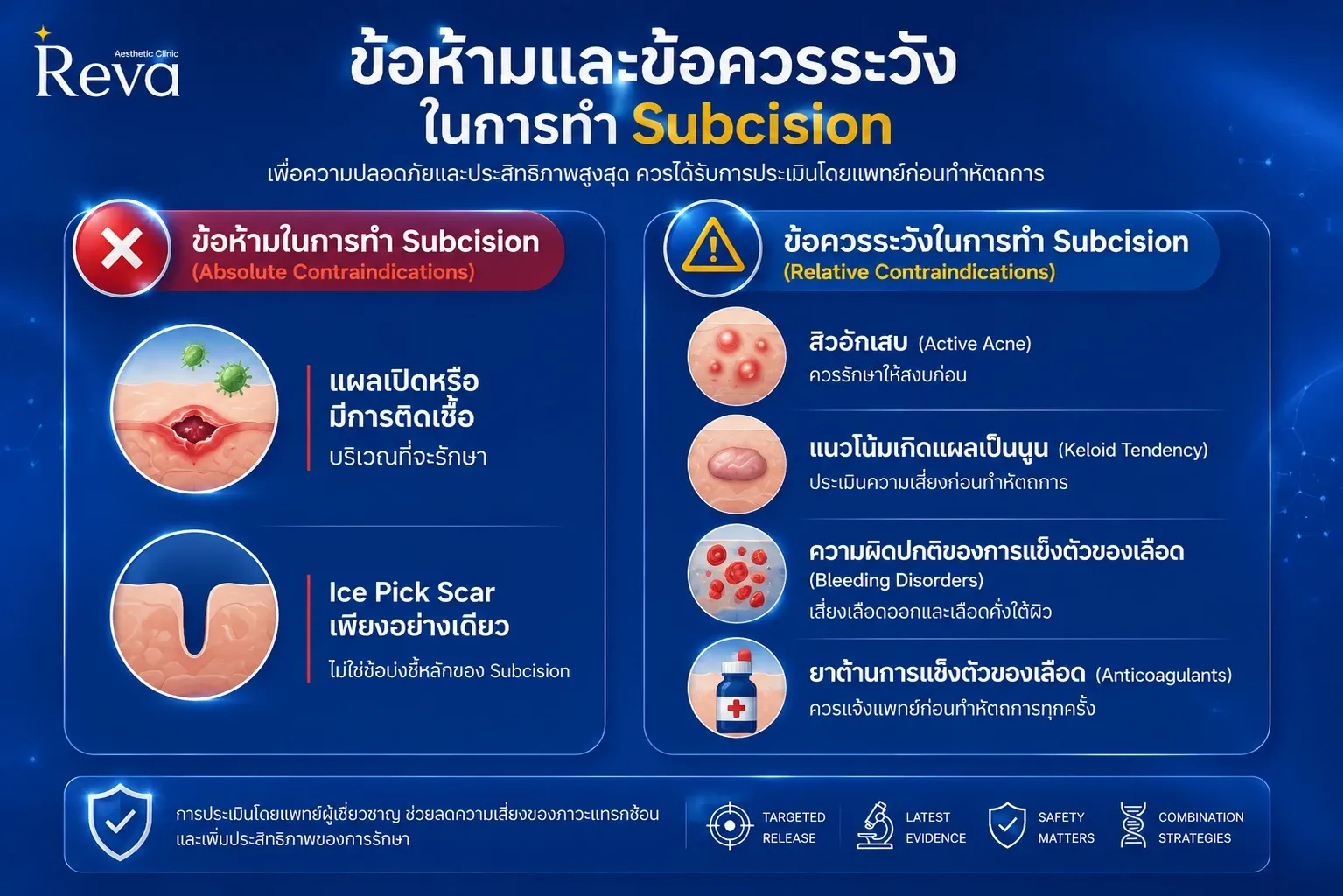
5. Contraindications and Precautions for Subcision
Although subcision is generally considered a safe and effective procedure when performed by an experienced physician, it is not suitable for every patient. Careful patient selection and pre-treatment assessment are essential to minimize complications and optimize treatment outcomes.
Absolute Contraindications
Subcision should not be performed under the following conditions until the underlying problem has been adequately treated.
Open Wounds or Active Skin Infection
Subcision should be avoided in areas with open wounds or active skin infections, as the procedure may increase the risk of bacterial spread, inflammation, delayed wound healing, and post-procedural complications.
Treatment should be postponed until the infection has completely resolved.
Isolated Ice Pick Scars
Subcision is not indicated for isolated ice pick scars because these scars result primarily from vertical tissue loss rather than fibrous tethering.
Since there are few or no fibrotic bands to release, subcision provides little clinical benefit. More appropriate treatment options include:
TCA CROSS
Punch Excision
Punch Elevation (selected cases)
Relative Contraindications
The following conditions are not absolute contraindications, but they require careful evaluation and individualized treatment planning before performing subcision.
Active Inflammatory Acne
Patients with active inflammatory acne should have their acne adequately controlled before undergoing subcision. Performing the procedure in the presence of active inflammation may increase the risk of infection and new scar formation.
History of Keloid or Hypertrophic Scarring
Patients with a personal history of keloids or hypertrophic scars may have a higher risk of abnormal scar formation following the procedure. A thorough risk assessment should be performed before treatment.
Bleeding Disorders
Patients with inherited or acquired bleeding disorders have an increased risk of excessive bleeding, bruising, and hematoma formation following subcision.
Anticoagulant or Antiplatelet Therapy
Patients taking anticoagulants or antiplatelet medications should inform their physician before treatment. The risks and benefits should be carefully assessed on an individual basis, and patients should never discontinue prescribed medications without consulting the prescribing physician.
Clinical Pearl
Successful subcision begins with appropriate patient selection. Careful evaluation of scar type, skin condition, medical history, and current medications is just as important as selecting the proper technique or instrument. Proper patient selection not only improves treatment outcomes but also minimizes the risk of complications.
6. What Instruments Are Used for Subcision?
Several instruments can be used to perform subcision, each with unique advantages, limitations, and clinical indications. Today, subcision devices are generally classified into three main categories:
Sharp Needles
Blunt Cannulas
Energy-Assisted Devices
No single instrument is ideal for every patient. The choice depends on scar morphology, the severity of fibrous tethering, treatment location, and the physician's experience and technique.
1. Sharp Needle
The sharp needle is the traditional instrument used for subcision. Its cutting tip allows the physician to efficiently release dense fibrous tethering beneath the skin, making it particularly effective for scars with strong fibrotic attachments.
Advantages
Excellent at releasing dense fibrous bands
Ideal for scars with severe tethering
Provides strong mechanical release
Limitations
Higher risk of bruising
Increased risk of hematoma formation
Requires precise control and extensive operator experience
2. Blunt Cannula
Blunt cannulas have become increasingly popular in recent years because they generally provide a safer treatment experience, particularly in areas containing major blood vessels and critical facial structures.
Instead of sharply cutting tissue, the rounded tip separates tissue planes while minimizing injury to surrounding structures.
Advantages
Lower risk of vascular injury
Less bruising and hematoma
Reduced postoperative pain and swelling
Higher patient satisfaction reported in several clinical studies
Limitations
Dense fibrous scars may require greater mechanical force to achieve adequate release
Cannulas with poor flexibility or lower elasticity may reduce treatment efficiency
3. Energy-Assisted Subcision
Recent advances have introduced energy-assisted subcision, combining mechanical release with thermal energy to simultaneously disrupt fibrotic bands and stimulate collagen remodeling.
Examples include:
Diode Fiber Laser
Radiofrequency (RF)-Assisted Cannula or Fiber
The concept behind this technique is to use controlled thermal energy to loosen fibrous attachments while promoting neocollagenesis, potentially reducing the amount of mechanical tissue trauma required.
However, current clinical evidence remains limited, and additional comparative studies are needed before these technologies can be considered standard alternatives to conventional subcision.
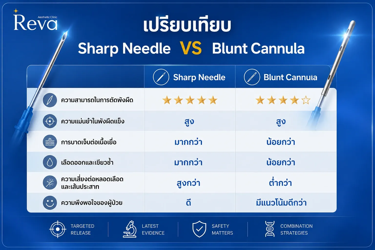
7. Sharp Needle vs. Blunt Cannula: Which Is Better?
Both sharp needles and blunt cannulas are effective instruments for performing subcision. At present, there is no definitive evidence demonstrating that one instrument is universally superior for all patients.
The optimal choice depends on several factors, including:
Acne scar type
Degree of fibrous tethering
Treatment location
Physician experience and preferred technique
More importantly, clinical outcomes are influenced far more by the physician's technique and anatomical understanding than by the choice of instrument alone.
Clinical Pearl
The best instrument is not the one with the sharpest tip or the bluntest cannula—it is the one selected appropriately for the patient's scar characteristics and used with precise anatomical technique.
Sharp Needle
Sharp needles are particularly effective for releasing dense or heavily tethered fibrous bands beneath the skin. Their cutting tip provides efficient mechanical release, making them a suitable option for scars with significant fibrotic attachments.
However, because they cut through tissue directly, sharp needles are generally associated with a higher risk of bleeding, bruising, hematoma, and soft tissue trauma compared with blunt cannulas.
Blunt Cannula
Unlike sharp needles, blunt cannulas separate tissue planes rather than cutting through them. This approach helps reduce the risk of injury to blood vessels and nerves, resulting in less bruising, swelling, and postoperative discomfort.
Several studies have also reported higher patient satisfaction with blunt cannulas, largely due to their improved safety profile and shorter recovery time.
Current Clinical Trend
Recent trends increasingly favor the use of blunt cannulas, particularly within the concept of Precision Subcision. This modern approach focuses on releasing only the pathological fibrous tethering while minimizing unnecessary tissue trauma and preserving normal facial support structures whenever possible.
Clinical Pearl
The goal of modern subcision is not simply to release scar tissue, but to do so as precisely and atraumatically as possible. Blunt cannulas have therefore become an increasingly popular choice for physicians aiming to maximize safety while maintaining effective scar release.
8. Potential Complications of Subcision
When performed by an experienced physician, subcision is generally considered a safe procedure. However, like any medical treatment, it carries the risk of side effects and complications. Fortunately, most adverse effects are temporary, self-limiting, and resolve with appropriate postoperative care.
Common Side Effects
Pain
Mild pain or tenderness at the treatment site is common during the first 1–3 days after subcision. Symptoms usually improve spontaneously or can be managed with prescribed analgesics.
Swelling
Swelling is an expected part of the normal healing process. It is typically most noticeable during the first 24–72 hours and gradually subsides within 1–2 weeks.
Bruising
Bruising occurs due to injury of small blood vessels beneath the skin and is one of the most common side effects of subcision, particularly when sharp needles are used. Most bruising resolves spontaneously within 1–2 weeks.
Uncommon Complications
Hematoma
A hematoma results from excessive bleeding beneath the skin and may present as a painful swelling or firm lump accompanied by more extensive bruising. Large or progressively enlarging hematomas should be evaluated by a physician.
Infection
Although uncommon, infection should be suspected if patients develop increasing pain, redness, warmth, swelling, purulent discharge, or fever after treatment. Prompt medical evaluation and antibiotic therapy may be required.
Hypertrophic Scar
Hypertrophic scarring is uncommon but may occur in patients with a predisposition to abnormal scar formation or when treating high-risk anatomical areas such as the perioral, perinasal, or periorbital regions.
Subcutaneous Nodule
Some patients may develop a small, firm nodule beneath the skin following subcision. In most cases, these nodules gradually resolve over several months. Persistent nodules lasting longer than six months should be reassessed by the treating physician.
Acneform Cyst
Rarely, acne-like cysts may develop at the treatment site. These lesions are generally manageable with medication or minor procedures, depending on their severity.
Subcutaneous Sinus Tract
Subcutaneous sinus tract formation is an uncommon complication characterized by an abnormal channel developing beneath the skin. Patients with this condition typically require further evaluation and appropriate treatment.
Rare Complication
Facial Nerve Injury
Facial nerve injury is extremely rare but remains one of the most important potential complications of subcision. Because branches of the facial nerve course through multiple anatomical regions of the face, meticulous knowledge of facial anatomy, proper instrument selection, and correct treatment depth are essential for minimizing this risk.
How to Minimize the Risk of Complications
Although serious complications are uncommon, their risk can be further reduced by following several key principles:
Carefully assess the type and severity of acne scars before treatment.
Select the most appropriate instrument and technique for each individual patient.
Follow the principles of Precision Subcision to minimize unnecessary tissue trauma.
Adhere strictly to all post-procedure care instructions.
Seek treatment from an experienced physician with a thorough understanding of facial anatomy.
Clinical Pearl
Most complications associated with subcision are technique-dependent rather than procedure-dependent. Careful patient selection, precise anatomical knowledge, meticulous surgical technique, and appropriate postoperative care are the keys to achieving safe and predictable outcomes.
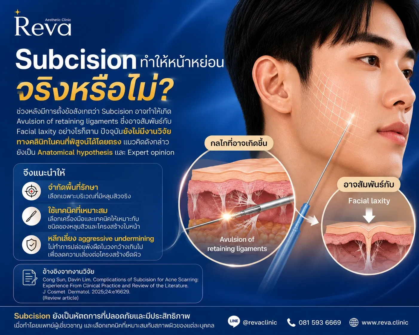
9. Can Subcision Cause Facial Sagging?
In recent years, concerns have emerged regarding whether aggressive subcision may increase the risk of facial laxity by causing avulsion of the retaining ligaments—the supportive fibrous structures that help maintain facial contour.
From a theoretical perspective, excessive tissue release could potentially disrupt these important anatomical structures. However, there is currently no clinical evidence in humans demonstrating that subcision directly causes facial sagging.
At present, this concept remains an anatomical hypothesis supported primarily by expert opinion, rather than by high-quality clinical studies.
Why Has This Become a Concern?
Retaining ligaments are key anatomical structures that anchor the skin, subcutaneous fat, and superficial soft tissues to the deeper facial framework. They play an essential role in maintaining facial contour and resisting the effects of gravity.
The concern arises from the theoretical possibility that extensive subcision, particularly when performed over a wide area or at an inappropriate tissue plane, may inadvertently disrupt these supporting ligaments.
If this occurs, it could theoretically reduce soft tissue support and contribute to facial laxity over time. However, this hypothesis has not been confirmed in clinical studies involving human patients, and no direct causal relationship has been established.
Current Evidence
Although aggressive subcision has been hypothesized to injure retaining ligaments, no human clinical study has demonstrated that subcision directly causes facial sagging or long-term facial laxity. Current concerns remain theoretical and should be interpreted with appropriate clinical context.The Treatment Philosophy Has Changed in 2026
Traditional subcision techniques often emphasized extensive undermining, aiming to release as many fibrous attachments as possible in an effort to maximize scar improvement.
Today, however, the approach has evolved toward Precision Subcision—a more targeted and anatomy-conscious technique that focuses on releasing only the pathological fibrous bands responsible for scar tethering while preserving normal tissue whenever possible.
Key Principles of Precision Subcision
Release only the fibrous bands responsible for acne scar tethering
Limit treatment to the affected areas (Targeted Release)
Avoid unnecessary extensive undermining
Preserve retaining ligaments and normal facial support structures whenever possible
This modern approach aims to minimize tissue trauma, reduce the risk of hematoma, shorten recovery time, and decrease the likelihood of long-term complications.
Precision Matters More Than Extensive Release
Today, successful subcision is no longer measured by how much tissue is released, but by how accurately the pathological fibrous tethering is identified and treated.
Excessively aggressive release may increase the risk of bleeding, hematoma, nerve injury, and prolonged recovery. It may also lead to iatrogenic scarring or unnecessary disruption of the normal supporting structures of the face.
For this reason, the physician's technique and understanding of facial anatomy are just as important as the procedure itself. Careful patient assessment, appropriate instrument selection, proper treatment depth, and precise release of pathological tethering are the cornerstones of modern Precision Subcision.
Clinical Pearl
Modern subcision is no longer about releasing more—it is about releasing smarter. Precision, anatomical knowledge, and tissue preservation have become the defining principles of contemporary acne scar treatment.
The Treatment Philosophy Has Changed in 2026
Traditional subcision techniques often emphasized extensive undermining, aiming to release as many fibrous attachments as possible in an effort to maximize scar improvement.
Today, however, the approach has evolved toward Precision Subcision—a more targeted and anatomy-conscious technique that focuses on releasing only the pathological fibrous bands responsible for scar tethering while preserving normal tissue whenever possible.
Key Principles of Precision Subcision
Release only the fibrous bands responsible for acne scar tethering
Limit treatment to the affected areas (Targeted Release)
Avoid unnecessary extensive undermining
Preserve retaining ligaments and normal facial support structures whenever possible
This modern approach aims to minimize tissue trauma, reduce the risk of hematoma, shorten recovery time, and decrease the likelihood of long-term complications.
Precision Matters More Than Extensive Release
Today, successful subcision is no longer measured by how much tissue is released, but by how accurately the pathological fibrous tethering is identified and treated.
Excessively aggressive release may increase the risk of bleeding, hematoma, nerve injury, and prolonged recovery. It may also lead to iatrogenic scarring or unnecessary disruption of the normal supporting structures of the face.
For this reason, the physician's technique and understanding of facial anatomy are just as important as the procedure itself. Careful patient assessment, appropriate instrument selection, proper treatment depth, and precise release of pathological tethering are the cornerstones of modern Precision Subcision.
Clinical Pearl
Modern subcision is no longer about releasing more—it is about releasing smarter. Precision, anatomical knowledge, and tissue preservation have become the defining principles of contemporary acne scar treatment.
10. What Should Be Done After Subcision to Prevent Re-tethering?
One of the primary goals of subcision is to release the fibrous bands that tether depressed acne scars. Once these fibrotic attachments are released, the skin can elevate naturally.
However, during the healing process, newly formed scar tissue may reconnect the skin to the underlying tissue—a phenomenon known as re-tethering. If this occurs, the acne scar may become depressed again, reducing the long-term effectiveness of treatment.
To minimize this risk, physicians may combine subcision with injectable materials that provide structural support beneath the scar, reduce the likelihood of re-tethering, and promote long-term tissue remodeling.
Each injectable material has a different mechanism of action, level of clinical evidence, and ideal indication.
Cross-linked Hyaluronic Acid (HA): The Most Evidence-Based Option
Among currently available injectable materials, cross-linked hyaluronic acid (HA) fillers have the strongest clinical evidence supporting their use in combination with subcision.
Immediately after fibrous release, HA filler can occupy the newly created space beneath the scar, providing structural support that helps prevent the skin from re-adhering to the underlying tissue.
This makes HA filler particularly suitable for acne scars associated with volume loss, including:
Rolling scars
Selected Boxcar scars
Depressed scars that elevate after subcision
Advantages of Cross-linked HA
Immediate structural support
Instant improvement in scar depression
Helps reduce re-tethering
Adjustable volume for different anatomical areas
Supported by the largest body of clinical evidence
However, HA fillers are not appropriate for every acne scar. Deep ice pick scars or scars with rigid fibrotic walls often require alternative treatments such as TCA CROSS, punch techniques, or laser resurfacing.
Hybrid Cooperative Complex HA (HCC-HA): A Tissue Remodeling Approach
Hybrid Cooperative Complex Hyaluronic Acid (HCC-HA) is not a dermal filler.
Instead, it is a unique hyaluronic acid technology that combines high- and low-molecular-weight HA using a thermal stabilization process. Rather than creating localized volume, HCC-HA diffuses throughout the tissue, supporting extracellular matrix remodeling and improving overall skin quality.
Unlike traditional fillers, HCC-HA focuses on biological tissue regeneration rather than immediate structural augmentation.
Clinical Study: HCC-HA vs. Cross-linked HA After Subcision
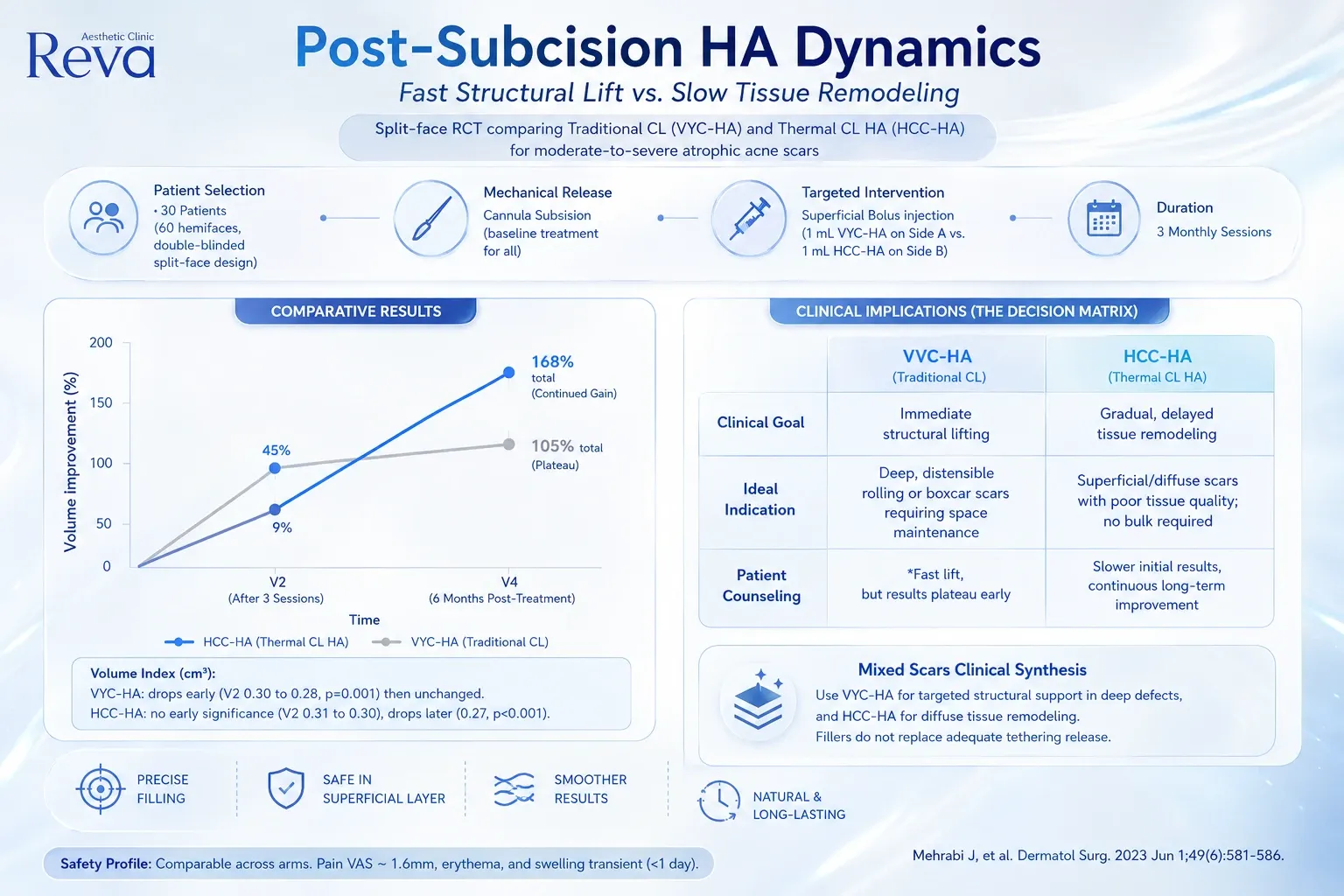
A prospective, randomized, double-blind, split-face clinical trial published by Mehrabi et al. (2023) evaluated 30 patients with moderate-to-severe atrophic acne scars.
All participants underwent subcision. One side of the face received cross-linked HA filler (VYC-HA), while the contralateral side was treated with Hybrid Cooperative Complex HA (HCC-HA).
Patients received three treatment sessions and were followed for six months.
Study Findings
Both treatments produced significant clinical improvement, but their treatment profiles differed substantially.
Cross-linked HA
Cross-linked HA demonstrated:
Rapid scar elevation
Immediate filling of the post-subcision space
Visible improvement shortly after treatment
It was particularly effective for:
Deep rolling scars
Boxcar scars associated with volume loss
Scars requiring immediate structural support
However, clinical improvement tended to plateau after treatment, with relatively limited additional improvement over time.
HCC-HA
HCC-HA produced a different response pattern.
Although the initial lifting effect was less pronounced than that of cross-linked HA, patients continued to show progressive improvement throughout the follow-up period.
The authors suggested that HCC-HA promotes tissue remodeling rather than simply replacing lost volume, allowing gradual improvement in skin quality and extracellular matrix architecture over time.
Structural Lift vs. Tissue Remodeling
The study highlights two fundamentally different treatment concepts.
Cross-linked HAHCC-HAProvides volume replacementPromotes tissue remodelingImmediate structural liftGradual biological regenerationRapid clinical improvementProgressive long-term improvementBest for deep depressed scarsBest for diffuse skin quality improvement
In simple terms:
Cross-linked HA lifts the scar.
HCC-HA restores the tissue.
Which One Should You Choose?
The answer depends on the characteristics of the acne scar, rather than the product itself.
Cross-linked HA is generally preferred for:
Rolling scars
Deep boxcar scars
Significant volume loss
Patients seeking immediate improvement
HCC-HA may be more suitable for:
Superficial atrophic scars
Thin or fragile skin
Poor skin quality
Patients seeking gradual collagen remodeling
Individuals who prefer treatment without traditional dermal fillers
A Simple Way to Understand the Difference
Think of cross-linked HA filler as providing structural support. It physically lifts depressed scars by occupying the space created after subcision.
In contrast, HCC-HA acts more like a biological skin regenerative treatment, gradually improving the extracellular matrix and promoting tissue remodeling without producing a pronounced volumizing effect.
Consequently, HA filler is generally more appropriate for localized structural defects, whereas HCC-HA may be better suited for diffuse skin irregularities and long-term tissue regeneration.
Although promising, clinical evidence supporting HCC-HA following subcision remains more limited than that for cross-linked HA fillers, and additional studies are needed to define its role more clearly.
Collagen Biostimulators After Subcision
Collagen biostimulators—including:
PLLA
PCL
PDLLA
Calcium Hydroxylapatite (CaHA)
have gained increasing attention for acne scar treatment because they stimulate the body's natural collagen production and improve dermal architecture over time.
Unlike HA fillers, these products do not provide immediate volume replacement. Instead, clinical improvement develops gradually through neocollagenesis.
Current evidence regarding their direct use for filling the post-subcision space remains limited. Differences in product formulation, dilution protocols, injection techniques, and treatment schedules make direct comparison between studies difficult.
As a result, there is currently insufficient evidence to conclude that collagen biostimulators can replace cross-linked HA fillers following subcision.
Nevertheless, they may be considered in carefully selected patients with:
Diffuse rolling scars
Generalized collagen loss
Age-related facial volume depletion
Thin or lax skin
Preference for gradual, natural-looking improvement
Proper product selection, injection depth, and technique remain critical.
PMMA: The Only FDA-Approved Filler for Certain Atrophic Acne Scars
In the United States, PMMA (Polymethylmethacrylate) is the only dermal filler with an FDA-approved indication for the correction of certain atrophic acne scars.
PMMA contains non-resorbable microspheres that provide long-lasting structural support while stimulating collagen production.
However, because PMMA is considered a permanent or semi-permanent filler, complications such as nodules, chronic inflammation, or granuloma formation can be more difficult to manage than with HA fillers.
Therefore, careful patient selection, meticulous injection technique, and experienced clinical judgment are essential. PMMA should not be considered the first-line treatment for every patient.
What About PRP and SVF?
Platelet-Rich Plasma (PRP) and Stromal Vascular Fraction (SVF) have also been investigated as adjunctive treatments following subcision.
Some studies suggest they may enhance wound healing, accelerate recovery, and improve collagen remodeling.
However, current evidence remains inconsistent due to significant variability in:
Preparation protocols
Concentration
Injection techniques
Treatment frequency
Outcome assessment methods
Consequently, standardized recommendations cannot yet be established.
Summary: Which Injectable Material Is Best After Subcision?
At present, cross-linked HA fillers remain the most evidence-supported injectable option following subcision, providing immediate structural support while helping reduce the risk of re-tethering.
HCC-HA represents a different treatment philosophy, focusing on biological tissue remodeling and gradual improvement in skin quality rather than immediate volumization.
Although collagen biostimulators such as PLLA, PCL, PDLLA, and CaHA show promising potential, current evidence for their routine use immediately after subcision remains limited.
Meanwhile, PMMA is the only filler with an FDA-approved indication for certain atrophic acne scars, but because of its long-lasting nature and limited reversibility, it should be used with careful patient selection.
Clinical Pearl
The key to successful post-subcision treatment is not choosing the newest injectable—it is selecting the right material based on scar type, scar depth, tissue tethering, volume loss, and each patient's individual treatment goals.
11. The Reva Clinical Perspective
Although subcision remains one of the cornerstone treatments for tethered atrophic acne scars, successful outcomes are not determined by how aggressively the fibrous bands are released. Instead, they depend on accurate scar assessment, appropriate instrument selection, meticulous technique, and a personalized treatment plan.
At Reva Aesthetic Clinic, we embrace the concept of Precision Subcision—a targeted, anatomy-guided approach that maximizes treatment efficacy while preserving healthy tissue and minimizing unnecessary trauma.
Our treatment philosophy includes:
Comprehensive assessment of each acne scar based on its morphology and anatomical location
Selection of the most appropriate instrument, whether a sharp needle or blunt cannula, according to the degree of fibrous tethering
Targeted release of only the pathological fibrous bands responsible for scar depression
Individualized Combination Therapy, incorporating HA fillers, Hybrid Cooperative Complex HA (HCC-HA), collagen biostimulators, laser resurfacing, or other regenerative treatments when clinically appropriate
Our goal extends beyond simply improving the appearance of acne scars. We strive to achieve safe, natural-looking, and long-lasting results while preserving normal facial anatomy and optimizing overall skin quality.
Dr. Songsak Suksantilap (Dr. Bank), M.D. Medical License No. 55316 Physician, Reva Aesthetic Clinic Updates on HCC for Acne Scar Treatment
Key Takeaways
✔ Subcision remains the gold standard for treating tethered rolling scars and selected boxcar scars.
✔ There is currently no clinical evidence demonstrating that subcision directly causes facial sagging or long-term facial laxity.
✔ Modern experts emphasize avoiding aggressive subcision and unnecessary extensive undermining, as excessive tissue release may increase tissue trauma and theoretically compromise important facial support structures.
✔ The treatment philosophy in 2026 has evolved toward Precision Subcision—releasing only the pathological fibrous tethering responsible for scar depression while preserving normal tissue, minimizing complications, and facilitating effective combination therapy.
✔ Successful acne scar treatment depends not only on the procedure itself but also on the physician's understanding of scar morphology, facial anatomy, proper instrument selection, and individualized treatment planning.
References
Alam M, et al. Subcision for Acne Scarring: Technique Update and Anatomical Considerations.Journal of Cosmetic Dermatology. 2025.
Goodman GJ, Baron JA. Postacne Scarring: A Quantitative Global Scarring Grading System.Dermatologic Surgery. 2006.
Jacob CI, Dover JS, Kaminer MS. Acne Scarring: A Classification System and Review of Treatment Options.Journal of the American Academy of Dermatology. 2001.
Mehrabi J, et al. Comparison of Two Hyaluronic Acid-Based Injectables Following Subcision for Atrophic Acne Scars: Structural Lift Versus Tissue Remodeling.Dermatologic Surgery. 2023.
Related Articles
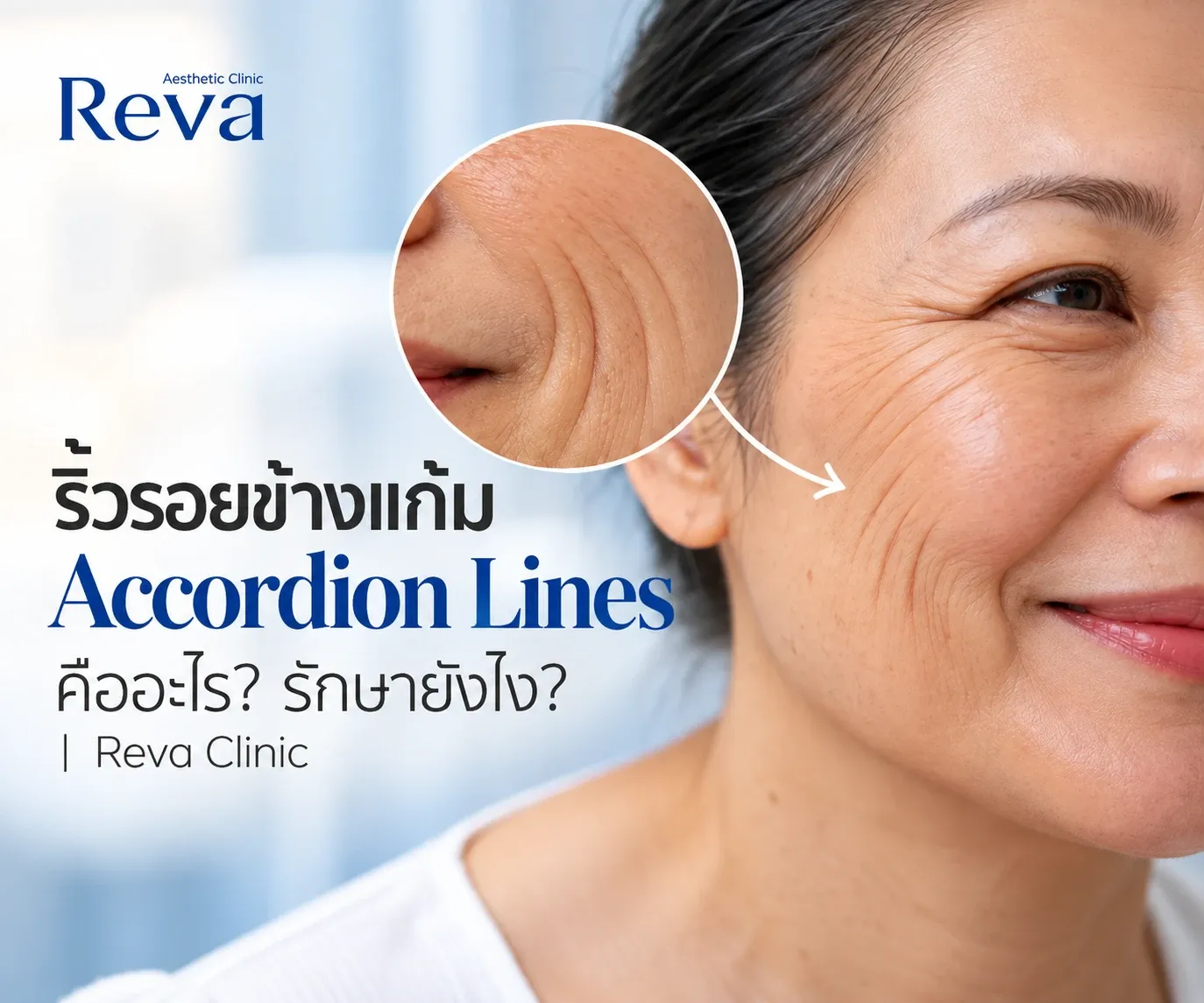
What Are Accordion Lines? Causes and Treatments for Cheek Wrinkles | Reva Clinic
Accordion lines are multiple cheek wrinkles that become more visible when smiling. Learn what causes them and how treatments differ for thin skin, skin laxity, and volume loss.
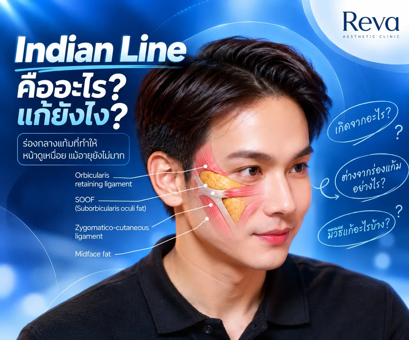
What Is an Indian Line? Causes and Treatment Options | Reva Clinic Meta Description
What is an Indian Line and why can it appear even at a young age? Learn how Indian Lines differ from nasolabial folds and explore treatment options including filler, collagen biostimulators and lifting treatments.
Dr. Bank of Reva Clinic Shares Filler Expertise at “Rheology Science of Multiphasic Filler: Lower Face Rejuvenation”
Dr. Songsak Suksantilapt (Medical License No. 55316), also known as “Dr. Bank” of Reva Clinic, shared his expertise in Rheology Science of Multiphasic Filler and Lower Face Rejuvenation at an Exclusive Hands-on Workshop on August 27, 2026. The session focused on facial assessment, filler rheology, and individualized treatment planning tailored to each patient’s facial structure and needs.
Program RedTouch Pro for Acne Scars: Does It Work and Which Scars Are Suitable? | Reva Clinic
Can Program RedTouch Pro improve acne scars? Learn how the 675 nm laser may help atrophic acne scars, which scar types may require combination treatment, and what recent Asian-skin research shows.
What Is Program Filler Teoxane? How Is It Different from Other Fillers and How Many Types Are There? | Reva Clinic
Program Filler Teoxane is a filler from Switzerland designed to deliver natural-looking results. Learn about the unique RHA technology, the differences between each model, and which areas they are suitable for, including under-eyes, nasolabial folds, lips, and wrinkles, with professional guidance from Reva Clinic.